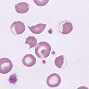
Eccentrocytes form under conditions of excess oxidant stress to the erythrocytes, which induces crosslinking of membrane proteins. Often, they are seen in association with Heinz bodies, which provide evidence of an oxidant effect on hemoglobin. Such cells have been seen in association with sydromes of oxidant injury in dogs, cats, and horses (e.g. onion and red maple leaf toxicity, glucose-6-phosphate dehydrogenase deficiency).
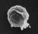
Note that when the thin membrane of the eccentrocyte is removed or ruptures, a small cell which lacks central pallor is formed. These spherocytic cells are termed "pyknocytes" and can be mistaken for spherocytes under light microscopy. Electron microscopy usually reveals the membrane "tags" (whereas spherocytes are uniformly spherical) as shown in the image on the left. |