|
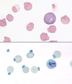
Blood from a cat with Heinz body hemolytic anemia associated with
acetaminophen toxicosis. Upper panel: the large Heinz bodies are causing
severe distortion of the cell outline. Note the eccentrocyte in the upper
left corner, and the ghost red cell with attached Heinz body in the upper right
corner. Lower panel: stained with New Methylene Blue.
|
Heinz bodies:
Oxidation of exposed sulfhydryl (SH) groups on hemoglobin causes formation of disulfide
bonds and distortion of the tertiary structure of the hemoglobin molecule. The result
is precipitation of hemoglobin, which may then coalesce to form intracellular
inclusions called Heinz bodies (HB). Small, non-pathologic "endogenous" HB can be found in blood
of many healthy cats, but their presence is abnormal in other species. Increased numbers of these "endogenous" HB are found in cats with diabetes mellitus (associated with ketone production), lymphoma and hyperthyroidism. Although many of these cats are not anemic, their red blood cell lifespans are reduced and they have lower hematocrits than cats with the same disease but lower numbers of HB.
Heinz bodies may be seen in Wright's stained smears, especially if they are large and distort the red cell membrane. In contrast, small "endogenous" HB are harder to visualize. HB can be easily observed with supravital stains, such as new methylene blue, as illustrated in the above photomicrograph.
Extensive HB formation and attachment to the membrane increase
the rigidity of the red cells and render them susceptible to fragmentation and entrapment
in the spleen where they are phagocytized. Heinz body hemolytic anemias are caused by oxidant injury from:
- Toxins: Wilted red maple leaves, Brassica species (kale, turnips), zinc, copper (sheep).
- Drugs: Acetaminophen, vitamin K, phenothiazine drenches, benzocaine.
- Inherited disorders: Enzyme or mineral deficiencies (necessary for enzyme function) that affect pathways that guard against oxidant injury, e.g. G-6-PD deficiency in horses, selenium deficiency in cattle.
Diagnosis of HB hemolytic anemia is based on
finding regenerative anemia with characteristic red cell morphologic abnormalities (HB, +/- eccentrocytes, keratocytes).
|